Researchers use AI to describe two biological subtypes of MS
Scientists at UCL have published research showing that people with MS can be split into two biological subtypes, based on data from brain images and blood tests.
You can be diagnosed with one of three types of MS: relapsing remitting, secondary progressive or primary progressive. These labels are used to guide treatment choices and help people understand the pattern of symptoms to expect.
But research has shown that these categories don’t always reflect the underlying biology of MS. For example, some people with progressive MS can experience relapses. This can make it more difficult to predict how someone's MS might progress and make sure people get the right treatment at the right time.
What did the researchers do?
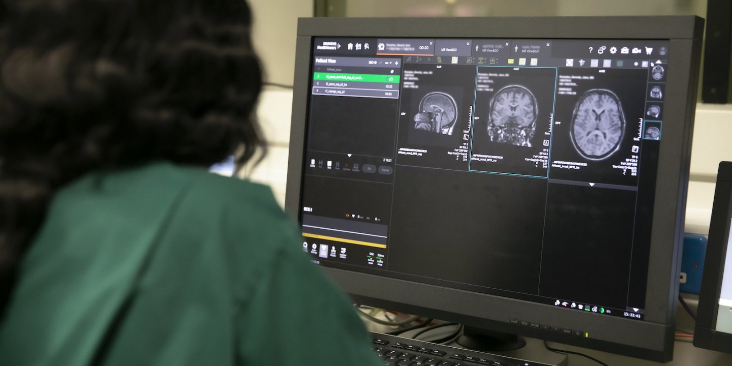
In this study, researchers used machine learning, a type of artificial intelligence (AI). Machine learning tools are trained to analyse huge datasets and recognise complex hidden patterns, better and faster than a human could.
This AI approach has been used with MRI data before. In 2021, Dr Arman Eshaghi and his team discovered their new machine learning tool could divide people with MS into three subtypes based on the type of damage in their brains. But MRI scans don’t capture everything that’s driving MS progression.
In this study, they used the same machine learning tool to combine MRI scans with information from blood tests to give us a more accurate picture. They looked at serum neurofilament light chain (sNfL), a protein that’s released into the blood when nerves are damaged. Higher levels of sNfL indicate more damage is taking place. They used existing data from 634 participants with relapsing remitting or secondary progressive MS.
What did they find?
By combining this data, researchers were able to identify two distinct subtypes of MS:
-
Some people had high levels of sNfL early in the condition. And MRI scans showed visible damage in a part of the brain called the corpus callosum. People in this group also developed new lesions more quickly. This suggests that their MS is more aggressive from the beginning.
-
In the second subtype, people had lower levels of sNfL to begin with. MRI scans showed brain shrinkage in areas of the brain called the limbic cortex and deep grey matter. And their sNfL levels rose later on. This suggests that their MS progresses more slowly and quietly, with more obvious damage occurring later on.
Dr Arman Eshaghi, who led the study, said:
“By using an AI model combined with a highly available blood marker with MRI, we have been able to show two clear biological patterns of MS for the first time. This will help clinicians understand where a person sits on the disease pathway and who may need closer monitoring or earlier, targeted treatment.”
What does this mean for people with MS?
MS is a complex condition, with many different biological processes likely driving it. Research like this could help doctors understand and more accurately describe someone’s individual MS. And allow researchers to develop new treatments that target this biology.
And because changes seen in MRI scans and blood tests often appear before any clinical changes in MS, using data-driven approaches like this could help clinicians to identify people who are at an increased risk of progression earlier.
Caitlin Astbury, our Senior Research Communications Manager, said:
“Over 150,000 people in the UK live with MS, with 135 diagnosed each week. This research adds to growing evidence supporting a move away from the existing descriptors of MS (like ‘relapsing’ and ‘progressive’ MS) and towards terms that reflect the underlying biology of the condition. This could help identify people at an increased risk of progression. And allow people to be offered more personalised treatment.”
But the researchers acknowledge there are barriers to bringing their research into everyday clinical care. For example, although MRI and sNfL are becoming more widely available, not all clinicians have access to the tools needed to do this analysis. And the way MRI scans are done often differs between hospitals. So they still need to do more research to overcome these hurdles.
You may also be interested in


Join the Research Network
Our Research Network make sure all the research we fund reflects the needs and interests of people living with MS. And they help us talk about it in a way that’s accessible to everyone.
Anyone who lives with MS or cares for someone who does can get involved. It doesn't matter where in the UK you live or how much research experience you have.
