Under the microscope: measuring myelin in people with MS
Dr Chris McMurran from Cambridge University tells us about the technology that can help us find new treatments to repair myelin.
There are lots of exciting therapies that work in the lab to boost the repair of myelin, the protective coating around our nerves. The next stage in stopping MS is getting these breakthrough discoveries to work safely and effectively on people.
Recently, I also made the jump from the lab to working with people with MS. I’m a junior doctor training to be a neurologist, and I’m helping to run clinical trials into myelin repair at the University of Cambridge.
There’s an important question to ask when planning any clinical trial - how do we know if our treatment is working?
Measuring symptoms and disability
If a drug can boost myelin repair, over time we hope that people with MS who take it will have fewer symptoms.
This sounds straightforward, but it might take years for the effect to be obvious. The treatment may be working but not enough to see an improvement in symptoms during a clinical trial that lasts only a few months. This means we might miss treatments that are working.
Measuring myelin
To get around this, we can measure myelin itself. In a lab I can look under a microscope and see myelin directly – but for studies with people we need to be a little more creative. Here are a few of the techniques we use to test whether myelin is being repaired by our treatments.
MRI
A type of MRI scan called magnetisation transfer ratio (MTR) has been developed that can see changes in the amount of myelin. It detects a signal that goes down when your myelin is damaged and back up as your myelin repairs. An increase in MTR signal from one scan to another tells us that myelin repair is happening.
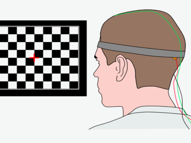
Visual evoked potentials (VEPs)
This technique measures how fast your nerves can carry a signal from your eye to your brain. We place wires over the skin on the ‘vision’ part of the brain. Then the person looks at an image – normally a flashing checkerboard pattern – and the wires pick up each signal as it arrives. The signals speed up when myelin is working better.
One downside is that VEPs only look in one part of the brain – and many people with MS don't have any problems with their vision. To get around this, there's work ongoing to use similar techniques that look at how fast other parts of the brain are working, such as moving an arm or feeling a pinprick.
Optical coherence tomography (OCT)
The retina (at the back of your eye) is the only part of your brain we can look at directly. Optical coherence tomography (OCT) makes a 3D picture of your retina – you’ve probably had this done at the opticians. From the 3D picture, we can see how many nerve cells are present.
This lets us measure the other main function of myelin – protecting nerve cells. If myelin repair happens, nerve cells are better protected from damage and we should see more of them in the picture.
Moving forward
It’s a very exciting time to be working in myelin repair research. Making these therapies a reality involves a team effort between scientists, clinicians and people with MS. Having the tools to measure myelin is a vital step towards this.
You may also be interested in


Will you help us stop MS?
We’ve now reached a crucial moment in our journey towards stopping MS.
You can help drive our research forward - so everyone with MS can get the treatment they need.
