MRI and MS: 7 things you need to know
MRI plays a vital role in how we diagnose and monitor MS. In fact, over 90% of people have their MS diagnosis confirmed by MRI.
1. What is MRI?
Magnetic resonance imaging, or MRI for short, uses strong magnetic fields to see inside the body. It’s particularly useful in MS as it allows us to measure what’s happening in the brain and spinal cord.
2. Why are MRI scans important for an MS diagnosis?
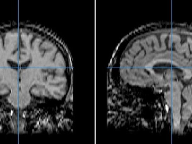
In MS your immune system attacks the myelin coating surrounding nerves. MRI scans can pick up these areas of damage, called lesions, in different parts of your central nervous system.
MRI has shaped how we monitor and treat MS too. It’s used to build a picture of how someone’s MS is changing over time. This can help to decide if a treatment is working.
Research has highlighted the benefits of regular MRI scans to monitor MS and inform treatment decisions. But, there's lots of factors to consider when deciding how often to have a scan. It's best to discuss this with healthcare professionals
3. What’s it like to have an MRI scan?
During an MRI scan you’ll be asked to lie flat on a bed that’s then moved inside the scanner. The scan usually lasts for 15 to 90 minutes, depending on how many images are being taken.
Although the procedure is painless, the machine itself can be very noisy. You’ll also be asked to keep as still as possible during the scan, so it’s important to get comfortable before it starts.
Read MS blogger Nicola's tips on having an MRI scan
4. Are there different types of MRI scans?
There are a number of different images that can be taken during a single MRI session.
A common type of MRI for MS is a T2-weighted scan, which detects all areas of myelin damage in the brain and spinal cord. Doctors may also use a type of scan called FLAIR, which makes it easier to spot the lesions.
Doctors will also use a contrast agent called gadolinium with a T1-weighted scan to focus on newer, active lesions. Gadolinium only highlights active damage because it can’t enter the brain unless there’s inflammation.
5. Can an MRI explain my symptoms?
What we can see on an MRI scan can explain the symptoms you might be experiencing. But it doesn’t always.
This is because many lesions may be in areas of the brain that don’t produce symptoms. And some areas of damage that could be causing symptoms might be too small to see on the scan.
6. How does MRI work?
MRI measures how much water there is in the body. Because different parts of the brain have different amounts of water, we can use MRI to distinguish them and build up pictures of the central nervous system.
The protective myelin coating is a fatty substance, so it repels water. This means we can measure how much myelin is present because it looks different to nerves and other cells in the brain on a scan.
7. What’s the future of MRI research?
Researchers sometimes rely on MRI scans in clinical trials to see the effect of a particular treatment. MRI scans can give us a window into the brain to see whether new treatments look promising much earlier on, before symptoms of progression even appear.
New, more powerful scanners will help us to detect even more subtle changes in the brain and spinal cord in MS. Improvements in MRI technology also mean we can run shorter trials to test the potential of new MS drugs, speeding up the whole development process.
MS diagnosis survey
We want to hear your experience of being diagnosed with MS. It doesn't matter when that was.
Answering our anonymous, short survey will help us shape the support we give people in the future.
The survey closes on Tuesday 30 June.

You may also be interested in