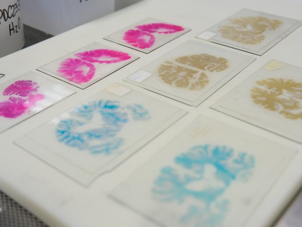
How brains donated decades ago change MS research today
Mia Humphries is a PhD student at Swansea University. We’ve committed to fund her work using brain tissue to find ways to predict how MS progresses. She tells us about her project.
I first came across MS in a neuroscience lecture. I then had the chance to visit my now-supervisor Dr Owain Howell’s lab and learn about his research.
I was fascinated to discover brain tissue donated after someone’s passed away, contains so much valuable information. It can reveal so much about the ongoing processes happening when you live with a condition like MS.
I’ve been in love with MS research ever since.
Working with brain tissue
We use tissue samples from the MS Society Tissue Bank in our research. Many of the brains we work with are from people who developed MS as far back as the 1970s, and we’re still using them to this day.
I’m so grateful for the people who donated and their families. Their brains have gone such a long way and told us so much already.
I know it’s such a big commitment, for someone to decide to donate their brain. There’s a lot to think about.
Understanding why MS progresses
The aim of my PhD is to identify what changes in brain tissue most strongly relate to disability progression.
Those are changes like:
- damage to axons (part of the nerve cell). Identifying this in brain tissue hasn’t actually been done much before. People have mostly focussed on spinal cord tissue, because the shape of the spinal cord makes it a bit more straight-forward
- the presence of certain inflammation-causing cells, like astrocytes and microglia.
- different types of lesions, like chronic active lesions (sometimes called smouldering lesions).
We have clinical information from doctors’ notes that come with the tissue samples. That’s things like what type of MS they lived with, how old they were when they were diagnosed and when they passed away.
So we can look for any patterns in this information.
Predicting progression in the clinic
The ultimate goal of our research is to develop a quick test that can predict whether someone is more likely to have worse progression. So doctors are able to offer more personalised treatment.
If you can understand exactly what’s happening in someone’s brain, you’ll know specifically what you need to target for that person with a treatment.
From brain tissue to blood test
It’s going to be quite a long journey. Once we’ve got an understanding of what changes in the brain tissue predict progression, we can move on to the CSF. CSF stands for cerebrospinal fluid – the liquid that surrounds the brain.
We can study the CSF collected from the same people who donated their brains. We’ll see if we can identify anything in the CSF that relates to the changes in brain tissue we found. Then we can check whether we see the same patterns in CSF samples from people living with MS now.
Finally, we’ll want to see if any of the markers we can spot in the CSF, can also be found in the blood. Because a blood test is much less invasive than taking samples of CSF from people, which needs a lumbar puncture.
Why tissue from people with MS matters
There are lots of methods for studying MS, and they all have benefits and drawbacks. For example, we’ve learnt a huge amount about MS from MRI scans. But you can’t tell the difference between certain types of lesion, or see axon loss, on an MRI scan. To see those cellular-level changes, you need the detail you get by looking at tissue from people.
And looking at tissue from brains donated so long ago, means we can really see what happens naturally in the brain when you have MS. Because there were no disease modifying therapies for MS when they were donated.
So I’m really excited to see where our project goes. I hope one day it’ll make a real difference to people with MS.
Vital research and resources like our Tissue Bank could hold the key to stopping MS for good.
Give today to support MS research.
You may be interested in


One donation. Double the impact.
We did it! Thanks to our match funders and your generous support, we've hit our £500,000 target!
But you can still help to raise money for vital MS research.
