Seeing MS differently: The role of eye scans in MS research
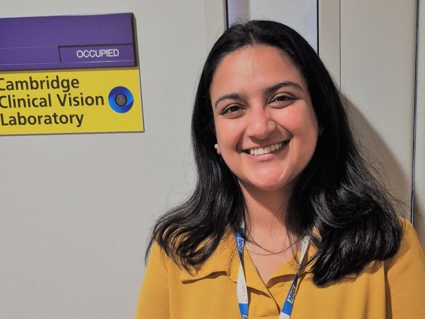
Riddhima Gautam is a Senior Ophthalmic Technician at the Cambridge Clinical Vision Laboratory. We spoke to Riddhima to find out more about her work and the role eyes play in MS research.
Can you tell us a bit about yourself?
My journey to working in research was a very unusual one. I'm originally from Nepal, but I did my undergraduate degree in India and worked there for a few years as an optometrist (a type of eye care professional who diagnoses and manages eye conditions).
But I always knew that I wanted to be involved in research. That’s why I moved to the UK in 2021 to pursue my master’s degree and specialise in eye research. In 2023, I moved to Cambridge where I now work in the Clinical Vision Lab – and I absolutely love it!
How is research into the eye connected to MS?
When you have MS, you tend to have inflammation of your optic nerve. And this type of inflammation can lead to the loss a type of cell called retinal ganglion cells. These ganglion cells are very important — they send signals from the eye to our brain and back.
Losing these cells can delay the transmission of these signals. This can impact how well you process images and cause disturbances in your vision.
Using eye scans allows us to see these changes. It's such a tiny organ, but you can get so much information from it. But eye research is also used in other conditions, like high blood pressure, diabetes, and different types of autoimmune disease.
What type of scans do you do?
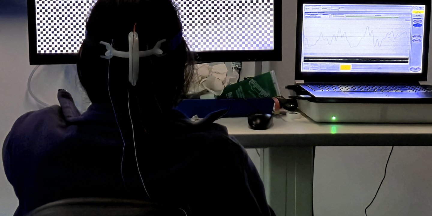
We use a lot of different types of cutting-edge eye scan techniques. One type is called the Visually Evoked Potential (VEP) test. During a VEP test, you look at a screen with a flashing checkerboard while small sensors on your head record your brain’s electrical response to what you see. These responses show how well visual signals travel from your eyes to the back of your brain. You can imagine the VEP test like a hearing test, but for your eyes and brain. It checks how quickly and clearly your brain reacts to what your eyes are seeing.
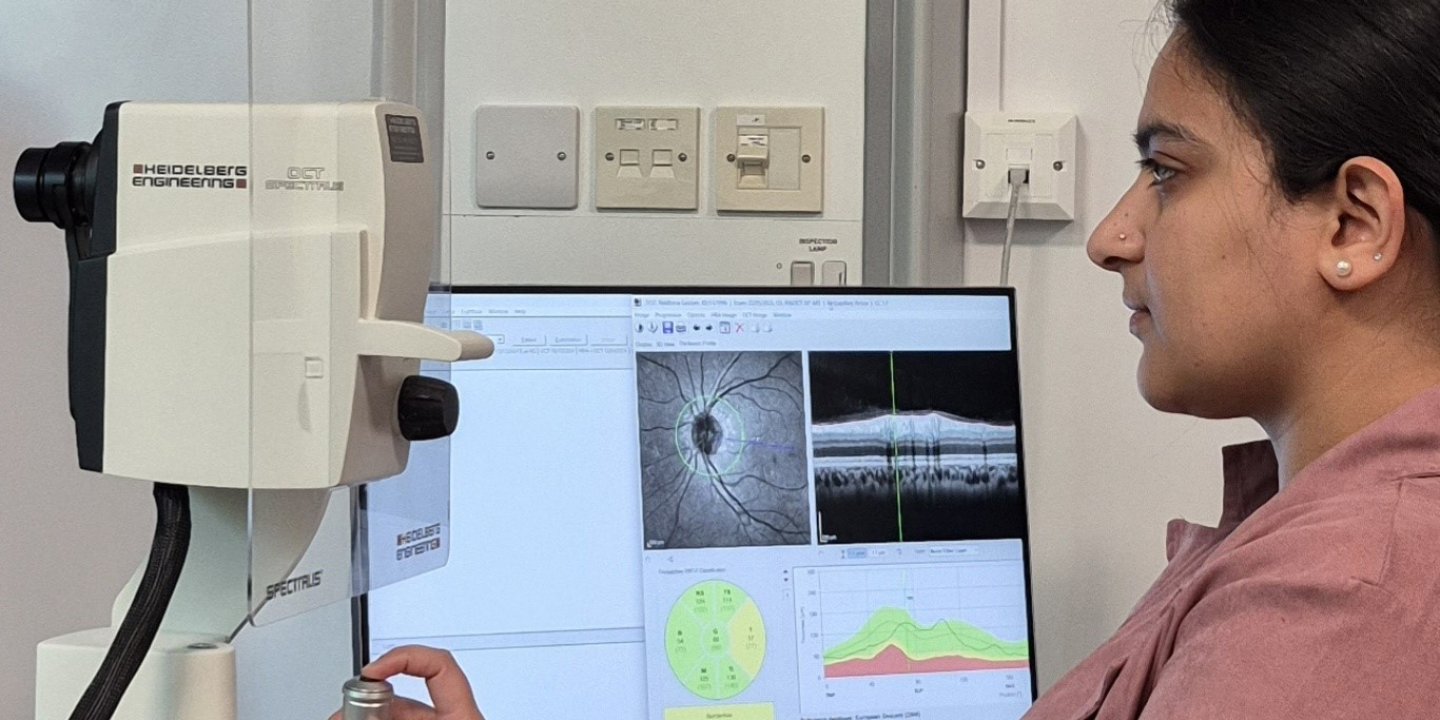
Another type of test is called Optical Coherence Tomography (OCT). It’s a non-invasive scan that tells us if there are any changes to your retina (tissue at the back of your eye) or optic nerve. Like the loss of ganglion cells I’ve mentioned earlier.
And we do a bunch of different tests to look at how well you can see colour and how sensitive your eyes are to different light contrasts.
Another very new technique in MS research is eye tracking. We use a camera to track different eye movements, which helps us to learn if someone’s MS has affected how well they can move their eyes.
What’s your favourite part of your job?
I love being able to come to this lab and make a difference to the lives of people by contributing to research. Ultimately, the techniques I’ve mentioned help us gather information about someone’s MS. And they can be used in research to make new discoveries or to test how well new treatments are working.
I learn something new every day. And I love to share that with patients. They’re often amazed by seeing their own scans and the information I can share with them. And I think that gives me a lot of satisfaction.
What’s your advice for people considering taking part in research?
My advice is to ask questions — to clinicians and researchers, but also to peers who are already taking part in research. There’s no harm in getting information and then it's completely up to you if you want to take part.
There are lots of different ways to get involved — for example, clinical trials, but also observational studies like the UK MS Register or taking part in Patient and Public Involvement (PPI) activities.
You may also be interested in


Will you help us stop MS?
We’ve now reached a crucial moment in our journey towards stopping MS.
You can help drive our research forward - so everyone with MS can get the treatment they need.
