
Spinal cord imaging in people with MS
MS is a condition that affects the brain and spinal cord. Nerve damage has mostly been studied in the brain. Even though nerve damage in the spinal cord has been strongly linked to disability, we know much less about it.
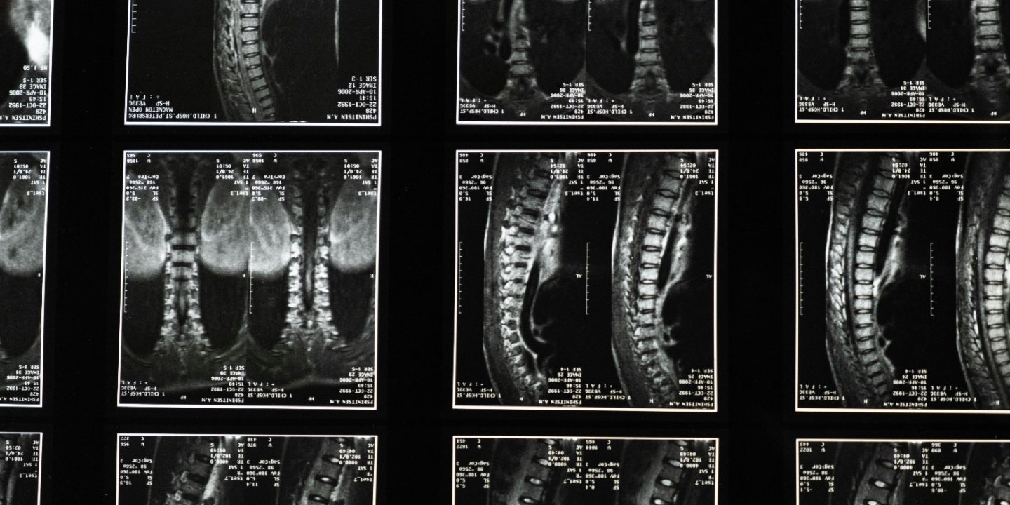
Current MRI scans can only show general damage to the spinal cord. We need better imaging techniques to be able to measure damage more accurately, especially in the early stages of the disease.
About the project
This project aims to develop imaging tools to measure nerve damage throughout the whole spinal cord in people with MS. Rozanna and the team will develop and test a novel MRI technique to precisely measure:
- How much tissue is lost over time (known as spinal cord atrophy).
- The extent of damage to the protective coating around the nerves (myelin).
- Any damage to the nerve fibres.
The team will first test this technique in healthy volunteers, followed by a study involving 20 people with early-stage MS. Participants will be scanned and will complete a questionnaire about their symptoms at the beginning of the study, and again six months later. If this project is successful, the team will then aim to carry out a larger study to test their technique in more people with MS.
How will it help people with MS?
This research could help us to understand more about the relationship between spinal cord damage and MS severity and progression. It could help with diagnosis and treatment decisions. And it could also provide a tool to accurately measure the effects of new treatments on the spinal cord in clinical trials.
You might also be interested in